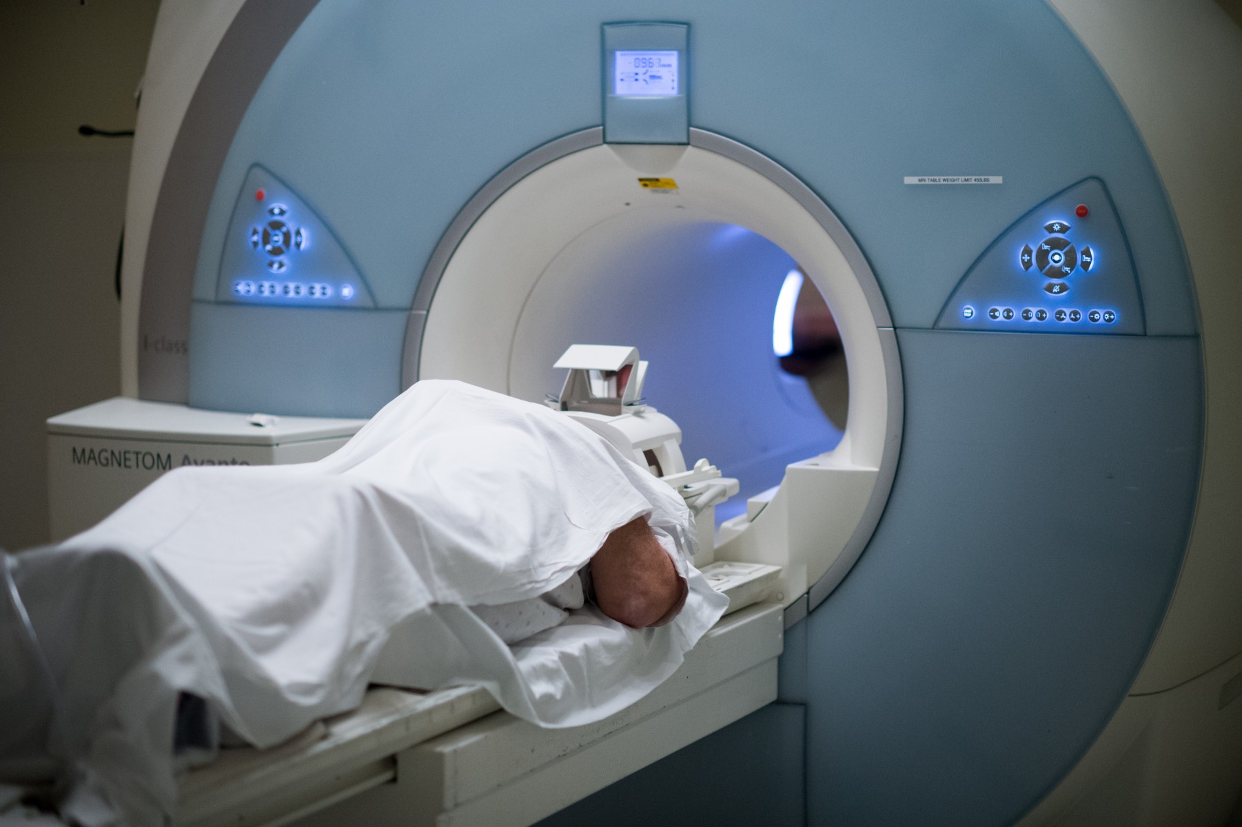
Tongue and perineural skull base disease better on MRI Neck, Skull base, Orbit Clinical Problem:ĬT defines ostial obstruction, bone changes MRI allows evaluation of bone marrow, CT for operative planning.ĭemyelination, Syrinx,Vascular abnormalities can be easily appreciated The disc, nerves and spinal cord can be seen clearly and in multiple planes.Ĭontrast essential to distinguish scar from recurrent disc prolapse after surgeryīenign Fracture v/s Pathological Fracture Images are helpful for identifying areas of scar tissue, abnormal development (dysplasia), and also changes in the brain white matter.Īdditional information is obtained like venogram and angiogram and that too without contrast.ĭynamic contrast evaluation helps in detecting small pituitary microadenomas Provides better details of brain structure than any other modality. MR spectroscopy- provides details about the metabolite contents of lesion ,which may help in differentiating infection from neoplasm. Multiplanar imaging-Better delineation of the extent and spread. Angiography provides additional information about the thrombosis of the vessel involved MRI detects infarcts in the hyperacute stage before it can be seen by any other modality. Spectroscopy,Diffusion tensor imaging,Breast MRI,Non contrast Angio,MRI Urography ,MRCP & Cardiac Imagingĭeep seated, microhemorrhages are detected. Advanced applications improves diagnosis.Ĭardiac CT, Denta Scan, Multiphasic Liver scan, Angiographies, 3 D imaging of bones,High resolution scan of thorax. Advanced applications can be done only on Multislice scanner.ġ.5 Tesla MRI-Higher magnetic field strength contributes high quality images, thinner slice thickness and potentially earlier and better detection of pathology. Multislice scanners offer reduced examination times and more importantly employ thin sections and improved resolution. No biological hazards have been reported with the use of the MRI. Magnetic resonance imaging (MRI) is a medical imaging technique most commonly used in radiology to visualize detailed internal structure of the body.,because of better soft tissue resolution.ĭespite being small, CT can pose the risk of irradiation.

The radiation is passed through the body and received by a detector and then integrated by a computer to obtain a cross sectional image that is displayed on the screen

AttributeĬT Scan or Computed tomography is a medical imaging obtained using X-rays. Rather than using radiation, this procedure exposes the body to a strong magnetic field, which affects the molecules in the body in such a way that a detailed picture can then be produced. MRI stands for Magnetic Resonance Imaging. To scan these tissues more effectively, a doctor will usually suggest an MRI scan. A CT scan is most beneficial for observing bone structure, and is less useful for scanning soft tissues in the body.


 0 kommentar(er)
